Neurosurgery
Neurosurgery for Brain Tumors
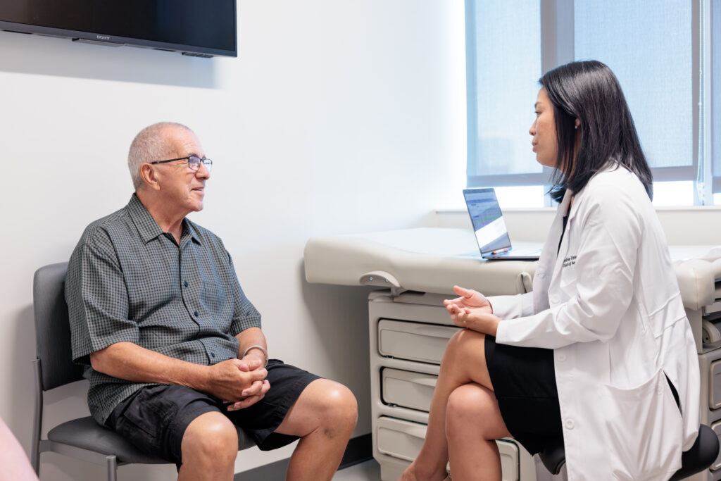
Neurosurgery for brain tumors refers to the surgical procedures performed by neurosurgeons to treat brain tumors. Brain surgery is often a crucial component of the overall treatment plan for patients diagnosed with brain tumors. The goals of neurosurgery for brain tumors include the removal of the tumor, relief of symptoms, preservation of neurological function, and improvement of the patient’s quality of life.
The world-renowned neurosurgeons at Barrow Neurological Institute perform more brain tumor surgeries than any other center in the United States. Utilizing the most advanced techniques and state-of-the-art equipment and technology, our neurosurgeons aim to remove the maximum amount of tumor tissue while preserving delicate areas of the brain.
Read on for a description of the key aspects of neurosurgery for brain tumors.

Tumor Resection
Tumor resection is the removal of brain tumor tissue. If a brain tumor is in a safe-to-reach area, the surgeon will try to remove as much as possible. Small tumors near accessible brain tissue can often be fully removed. However, if the tumor is close to sensitive areas or can’t be fully separated, surgery is riskier. In these cases, the goal is to remove as much as safely possible. Even partial removal can help with symptoms. Sometimes, only a small sample (biopsy) is taken to confirm the diagnosis.
Surgery to remove brain tumors has risks, such as infection, bleeding and stroke. Other risks depend on the tumor’s location in the brain. For example, surgery on a tumor near the part of the brain that controls movement (motor cortex) may cause weakness on one side of the body after surgery. Specific risks will be discussed prior to surgery during the consent process.
Craniotomy
“Crani” means bone, and “otomy” means temporarily removing and putting back. A craniotomy is a surgery that involves the removal of a piece of bone for a procedure and the return of that bone at the end of the surgery. Surgery to remove a brain tumor is an example of a surgery that often requires a craniotomy.
The patient is put to sleep and then positioned on an operating table with their head fixed on a headrest. A small amount of hair is clipped along the incision line. The skin where the incision will take place is washed to be sterile. An incision is then made on the skin, usually over the location of the tumor. After the skin is spread apart, the surgeon drills an opening in the skull (this is the craniotomy). The bone is removed and kept sterile until the end of the surgery. The surgeon then performs the specific procedures on the brain.
In the end, the bone is placed back and anchored with tiny, titanium screws and plates (the metal is not magnetic, cannot be felt through the skin, and does not set off metal detectors). The skin is closed, and the surgery is completed.
Awake Craniotomy
If a tumor is located near the area of the brain that controls motor movement or language function (eloquent cortex), the surgeon may recommend an awake craniotomy. The craniotomy portion (the temporary removal and return of bone) is the same as described in the craniotomy portion above. Once the brain is exposed, the patient is woken up (the “awake” portion of the surgery).
Once awake, the patient will be asked to follow commands by repeating words, naming objects or moving parts of their body. If the patient cannot follow commands when a specific area of the brain is stimulated, the surgeon knows that area of the brain is critical for function and will not remove it. Once language and motor function has been mapped out on the brain, the tumor is removed. The patient goes back to sleep while the remainder of the surgery is completed.
Asleep Mapping Craniotomy
If a tumor is located near the area of the brain that controls motor movement, the surgeon may recommend an asleep-mapping craniotomy. The craniotomy portion (the temporary removal and return of bone) is the same as described in the craniotomy portion above. The surgery starts with the standard steps of a skin incision, drilling of the bone and the temporary removal of the bone.
Once the brain is exposed, we stimulate areas of the brain while watching the patient’s body for any movement in the face, arms or legs. If the stimulation of a specific location in the brain results in movement, the surgeon knows that area of the brain is critical for movement and will not remove that area. If the stimulation of a specific area in the brain does not result in movement, the surgeon knows that area of the brain is safe to be removed. Once the motor function has been mapped out on the brain, the tumor is removed.
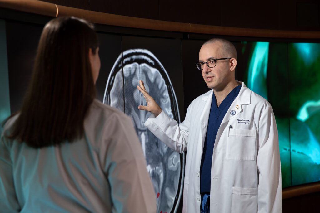
Intraoperative MRI (iMRI)
An intraoperative MRI (iMRI) is a state-of-the-art imaging technique used during brain tumor resections. Unlike traditional MRI scans, iMRI is performed in the operating room, providing real-time, high-resolution images of the brain during the surgery. This imaging allows surgeons to make precise decisions, ensuring thorough tumor removal while preserving healthy tissue. By integrating iMRI into brain tumor resections, surgeons can optimize the surgical strategy, leading to more accurate outcomes.
Functional Brain Mapping
Functional brain mapping is a technique used to identify and map specific functions, such as motor skills, language centers, and sensory perception, to precise regions of the brain. By stimulating various brain areas and observing the patient’s responses neurosurgeons can create a detailed map of the brain’s functional organization. This mapping helps ensure that critical areas are preserved and minimizes the risk of damaging essential functions to maintain the patient’s quality of life post-surgery. Functional brain mapping can be performed with the patient awake or asleep, depending on the functions needing to be mapped.
5-ALA Guided Surgery
Fluorescence-guided surgery using 5-aminolevulinic acid (5-ALA) is a surgical technique used by many neurosurgeons worldwide to treat gliomas. Patients drink the medicine 2-4 hours before surgery and the 5-ALA helps the neurosurgeon distinguish tumor tissue from normal adjacent brain during surgery. Your neurosurgeon may ask you to take this medication prior to surgery. Side effects are increased risk of sun sensitivity or sunburn with sun exposure. Patients should avoid direct sunlight for five days after surgery and wear sunglasses when exposed to sunlight.
STEREOTACTIC NEEDLE BIOPSY

In cases where there is a lesion (abnormal area) in the brain or spine with an unclear diagnosis, your neurosurgeon may recommend a biopsy. A biopsy is removing a small piece of tissue to send to the lab for pathology testing. This testing involves microscopic examination, staining with different dyes and molecular sequencing, and will help determine a diagnosis.
At Barrow, our neurosurgeons perform stereotactic needle biopsies. The biopsy needle is precisely guided by three-dimensional coordinates obtained from neuroimaging like MRI or CT scans. A small tissue sample is obtained using a specialized, navigated needle, which is inserted through a tiny skin incision and hole in the skull. This technique is particularly valuable for diagnosing brain tumors in challenging locations. Stereotactic biopsy offers patients a less invasive alternative to traditional open surgical biopsies, providing essential diagnostic information while minimizing risks and recovery time. The procedure is routinely performed outpatient, and patients can return home the same day after surgery.
Laser Interstitial Thermal Therapy (LITT)
LITT is a minimally invasive surgical technique performed in the operating room in which a thin laser fiber is stereotactically implanted directly into the brain tumor through a tiny skin incision and hole in the skull to thermally ablate tumor tissue in deep or difficult-to-access parts of the brain. The laser heats the tumor tissue to temperatures high enough to kill the tumor while preserving the surrounding normal brain tissue. The laser catheter is precisely guided by three-dimensional coordinates obtained from neuroimaging like MRI or CT scans. The treatment heat is monitored in real time using MR imaging. This procedure has a faster recovery time than open brain surgeries. The laser is completely removed after treatment.
MRI-Guided Focused Ultrasound (FUS)
Focused ultrasound (FUS) uses sound waves that are capable of disrupting the blood-brain barrier near and around the brain tumor to enhance drug delivery to the area. By disrupting the brain’s protective blood-brain barrier medications are more effectively able to get into the brain to kill the tumor cells. Your surgeon will place a stabilizing frame around your head which allows precise targeting of a location in the brain with the FUS. The MRI is used to precisely guide the delivery of focused ultrasound waves. This minimally invasive treatment option is used in combination with specific chemotherapies and is not applicable to all brain tumors currently.
Shunt Placement
A brain or spinal cord tumor can put pressure on the brain and increase the risk of hydrocephalus, which is a term that refers to too much water in the brain. “Hydro” means water, and “cephalus” means head. The water, in this case, is cerebrospinal fluid (CSF), and is stored in a small space in the brain’s center called the ventricles. A shunt placement is a standard treatment for hydrocephalus. The shunt is a thin (thinner than a pencil), flexible tubing with holes at the two ends. This tubing provides an alternative route for water to pass through and drain from the brain to another location in the body. The neurosurgeons at Barrow perform four types of shunt placement procedures.
Ventriculoperitoneal Shunt
A ventriculoperitoneal (VP) shunt travels from the ventricles (center of the brain) to the peritoneal cavity (abdomen). Specifically, it travels from the ventricles in the center of the brain to the surface of the skull bone, then behind the ear where a regulatory valve is located, down the neck, down the chest, and into the abdomen (all under the skin). The cerebrospinal fluid that passes into the abdomen is small and the body can absorb it naturally without any issue. The implanted valve is used to regulate the amount of cerebrospinal fluid flow, and its setting can be changed through the skin without further surgery.
Ventriculoatrial Shunt
A ventriculoatrial (VA) shunt travels from the ventricles (center of the brain) to the blood vessel next to the heart. Specifically, it travels from the ventricles in the center of the brain to the surface of the skull bone, then behind the ear where a regulatory valve is located, down the neck, and into the vein next to the heart (all under the skin). The small amount of cerebrospinal fluid from the brain passes into the heart and mixes in with the blood with no adverse effects. The implanted valve is used to regulate the amount of cerebrospinal fluid flow, and its setting can be changed through the skin without further surgery.
Ventriculopleural Shunt
A ventriculopleural (VPleural) shunt travels from the ventricles (center of the brain) to the pleural cavity (lung). Specifically, it travels from the ventricles in the center of the brain to the surface of the skull bone, then behind the ear where a regulatory valve is located, down the neck, down the chest, and into the cavity around the lung (all under the skin). The cerebrospinal fluid that passes into the pleural space is small and the body can absorb it naturally without any issue. The implanted valve is used to regulate the amount of cerebrospinal fluid flow, and its setting can be changed through the skin without further surgery.
Lumboperitoneal Shunt
A lumboperitoneal (LP) shunt travels from the spinal canal to the peritoneal cavity (abdomen). Specifically, it travels from the space around your nerves in your spine (which is surrounded by cerebrospinal fluid), around the flank (the side of your body below your ribs), to the abdomen (all under your skin). The cerebrospinal fluid that passes into the abdomen is small and the body can absorb it without any issue. There is an implanted valve that is used to regulate the amount of cerebrospinal fluid flow, and its setting can be changed through the skin without further surgery.
Barrow Brain Tumor Neurosurgeons
 Read Bio
Nader Sanai, MDDirector, Ivy Brain Tumor Center and Chief of Neurosurgical Oncology, Barrow Neurological Institute
 Read Bio
Michael T. Lawton, MDPresident and CEO, Barrow Neurological Institute Neurosurgeon
 Read Bio
Kris Smith, MDNeurosurgeon
 Read Bio
John Wanebo, MDNeurosurgeon
 Read Bio
Kaith Almefty, MDNeurosurgeon
 Read Bio
Zaman Mirzadeh, MD, PhDMetabolism Core Leader Neurosurgeon
 Read Bio
Randall Porter, MDNeurosurgeon
 Read Bio
Andrew Little, MDNeurosurgeon
 Read Bio
Steve Chang, MDNeurosurgeon
To request a second opinion from a Barrow neurosurgeon, call 602-406-3396.
Neurosurgery for brain tumors requires a highly skilled and specialized surgical team, including neurosurgeons, neuroanesthesiologists, and neurophysiologists, working together to provide the best possible outcome for the patient. After surgery, patients may undergo additional treatments such as radiation therapy, chemotherapy, or targeted therapies, depending on the specific type and characteristics of the tumor.