Neuroimaging
Neuroimaging for Brain Tumors
Neuroimaging for brain tumors refers to the use of various imaging techniques to visualize and diagnose tumors in the brain. These imaging methods are essential for accurately identifying the presence, location, size, and characteristics of brain tumors. Neuroimaging plays a crucial role in determining the most appropriate treatment plan and monitoring the tumor’s response to therapy.
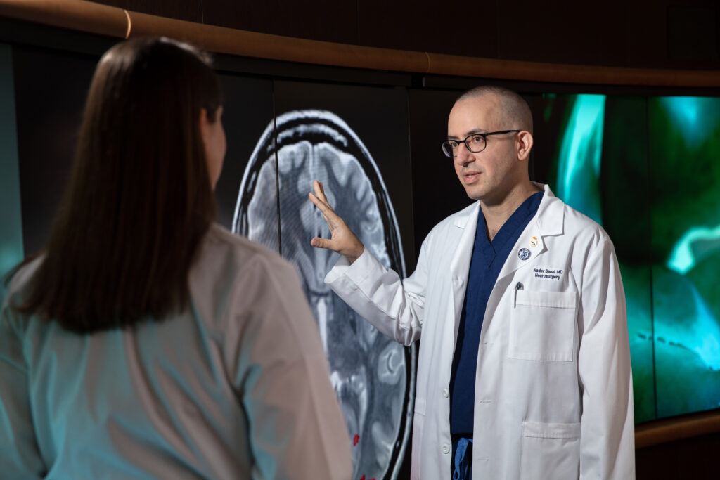
The Ivy Brain Tumor Center exists within Barrow Neurological Institute, where skilled neuroradiologists in Department of Neuroradiology at Barrow Neurological Institute collaborate closely with neuro-oncologists and neurosurgeons, employing high-resolution imaging methods to diagnose, strategize, and oversee the progress of patients with brain tumors.
Scroll down to learn about the most commonly used diagnostic and therapeutic imaging techniques used for brain tumors.
X-rays
X-rays are a type of medical imaging that uses a small dose of radiation to create pictures of the inside of the body. In brain tumor cases, X-rays are typically used to capture images of the skull and can help identify fractures, abnormalities, or conditions that might affect brain health including implanted devices such as shunts. While X-rays offer valuable insights into bone structures, they have limitations in visualizing soft tissues and intricate details, making them less commonly used for direct brain tumor evaluation.
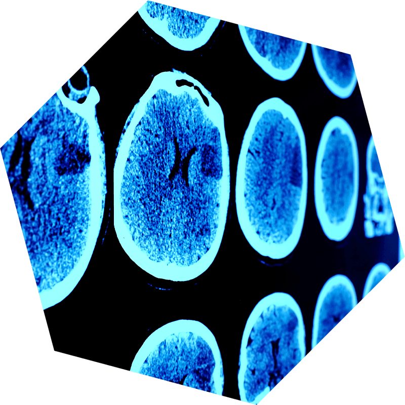
CT Scan
CT scan or computed axial tomography scan (CAT scan) is a painless test that uses X-rays (radiation) to produce the images. CT is an open machine; generally, claustrophobia is not an issue. CT is usually safe but does use radiation; therefore, your treating team monitors frequent exposure. In brain tumor diagnostics, CT scans provide clear images of the brain’s anatomy, aiding in detecting abnormalities, tumors, bleeding, and other conditions. By offering a rapid and comprehensive view, CT scans assist doctors in diagnosing brain tumors, assessing their size, location, and impact on surrounding tissues.
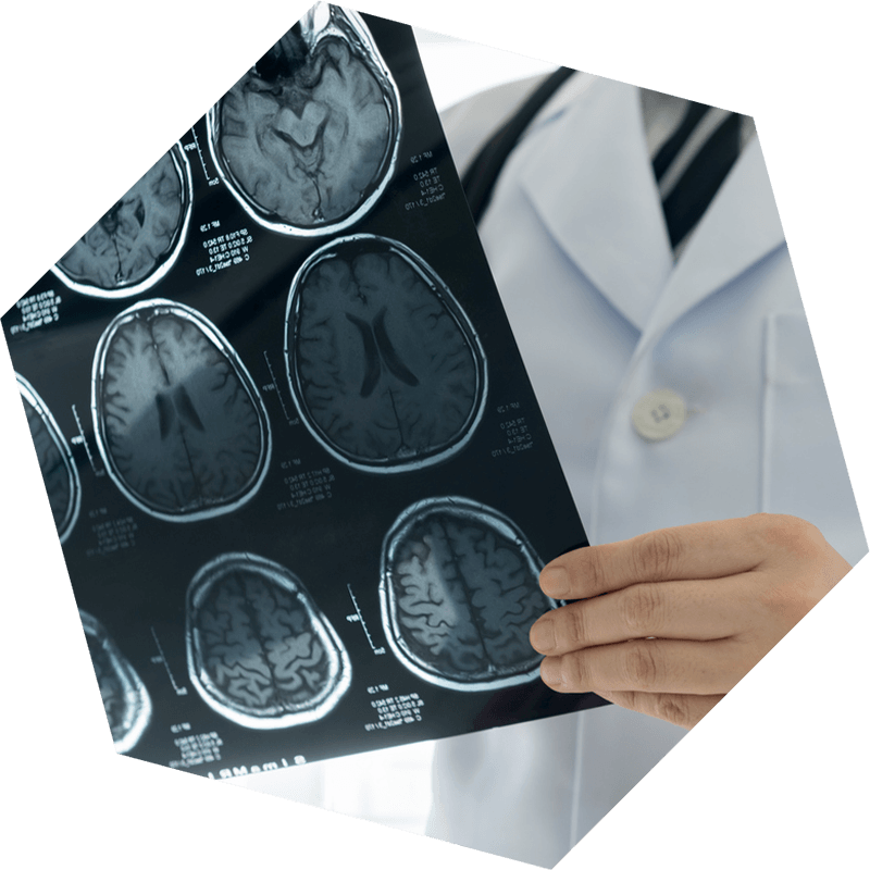
MRI
MRI stands for magnetic resonance imaging (MRI), a safe and painless test that uses a magnetic field to produce detailed images of the brain or spinal cord. Most brain tumor patients have regular MRIs ordered by their surgeon or oncology team. The frequency of MRIs for a patient depends on the type of tumor and the physician’s discretion. MRIs are generally safe and painless and use no radiation. The MRI machine is somewhat enclosed and can be challenging for patients with claustrophobia. In those cases, light or heavy sedation can be administered.
Other MRI techniques include MRI with and without contrast, perfusion MRI, MR spectroscopy, and functional MRI which can map out specific functions such as motor skills, language centers, and sensory perception.
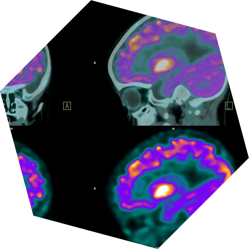
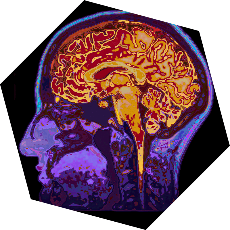
PET Scan
Positron Emission Tomography (PET) scans reveal how active tissues are by using a tiny bit of radioactive material. These scans detect gamma rays emitted when this material interacts with cells. In brain tumor imaging, PET scans help distinguish active tumor cells from healthy ones due to the higher metabolism of cancer cells. This aids in diagnosing aggressive tumors, tracking their spread, monitoring treatment effects, and planning treatment strategies. Working with other imaging methods, PET scans offer essential insights for understanding and managing brain tumors. In cases of brain metastases of cancer from other organs, or in staging for CMS lymphoma, your doctor may order a PET scan of your body.
Barrow Neuroradiologists


Neuroimaging plays a vital role in the diagnosis, treatment planning, and monitoring of brain tumors, enabling healthcare professionals to provide the most effective and personalized care for patients.